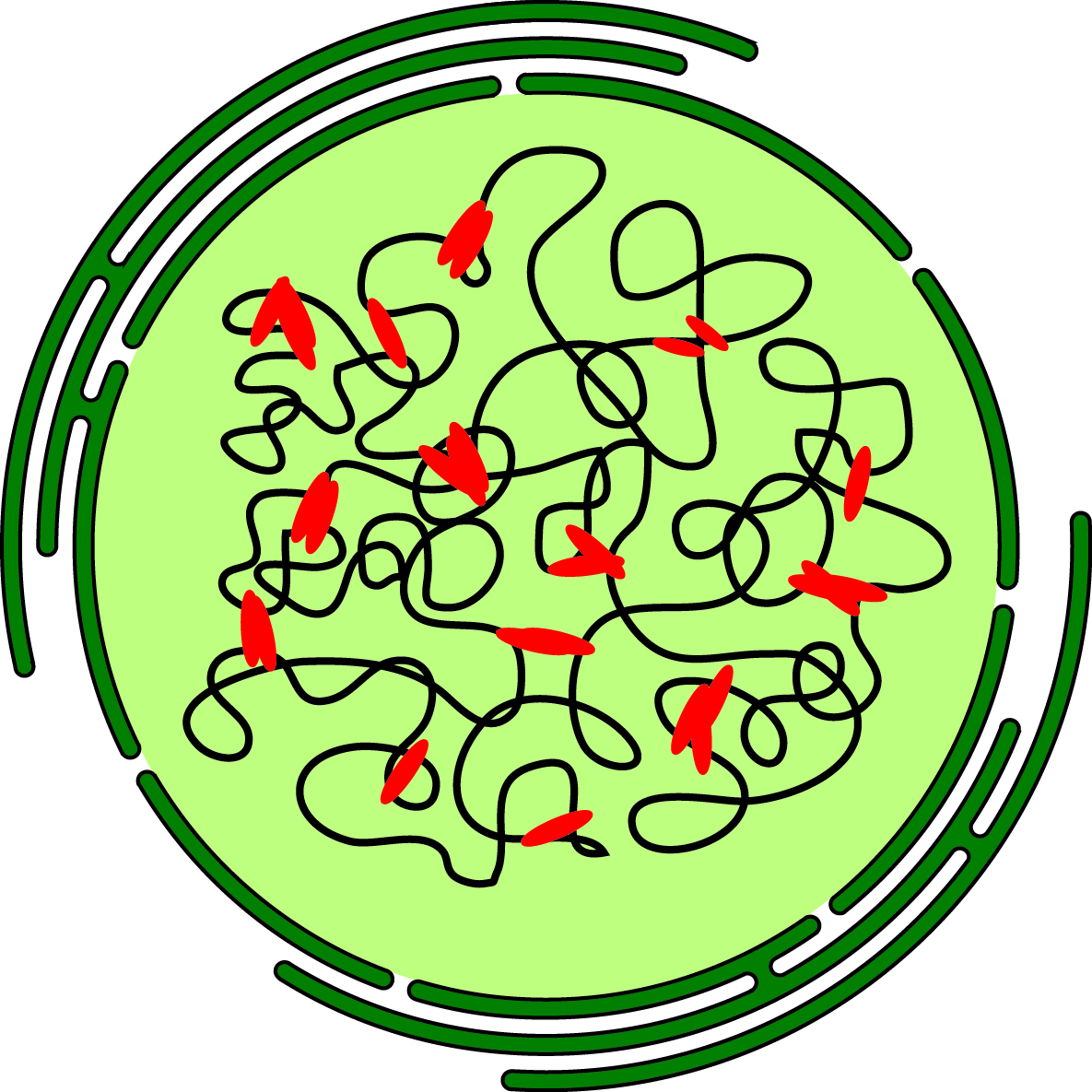

Single molecule mechanisms of spatio-temporal chromatin architecture
ERC Starting Grant - ChromArch

Chromatin packaging into the nucleus of eukaryotic cells is highly sophisticated. It not only serves to condense the genomic content into restricted space, but mainly to encode epigenetic traits ensuring temporally controlled and balanced transcription of genes and coordinated DNA replication and repair. Owing to fluorescence in situ hybridization (FISH) assays and novel chromatin conformation capture (3C) techniques, it became evident that regulatory traits are not only encrypted along the one-dimensional sequence of DNA, but within the three-dimensional arrangement of chromatin. This non-random chromatin organization ranging from the state of compaction by nucleosomes over topologically associated domains to the relative location of whole chromosomes is of utmost importance for the correct read-out and control of genetic information. Transcription of a gene for example can be significantly increased by long-range chromatin interactions between enhancers and promoters.
A rich variety of biomolecules involved in organizing the genome has been identified, including nuclear lamina, non-coding RNA molecules and architectural proteins. However, despite increasing interest in spatio-temporal chromatin organization, mechanistic details of their contributions to establishing and maintaining chromatin topology are not well understood. Many open question beyond the identification of participating factors are unanswered. How long does it take for the distal ends of a chromatin sequence to form a loop, how long do functional connections, for example enhancer-promoter interactions, persist and what are the molecular mechanisms of tethering two genetic regions together?
We aim at unveiling molecular mechanisms of chromatin organization by quantitative in vivo and in vitro single molecule experiments. 3C techniques can detect changes in chromatin organization upon cell differentiation or during the cell cycle, but important information on the frequency of interactions within a cell population and the temporal stability of distant chromatin associations remain elusive due to averaging over many cells and the destructive nature of these methods. We thus use single cell and single molecule fluorescence microscopy to measure the dynamics of organizational structures within chromatin and to study the molecular mechanisms of biomolecules mediating chromatin topology in the nucleus. In complementary single molecule force spectroscopy experiments we study mechanisms of chromatin structure formation in vitro. Our goal is to enhance the mechanistic understanding of three-dimensional chromatin architecture and its regulatory effects on nuclear functions and to inspire experiments on the potential therapeutic utility of controlled modification of regulatory traits mediating chromatin topology.
