Ältere Mitteilungen
We congratulate Clarissa Read and Kavitha Shaga Devan for the successful raising of a grant of the Baden-Württemberg-Stiftung “Methoden der Lebenswissenschaften” in cooperation with Timo Ropinski.
We congratulate Ulrich Rupp for a poster prize at MC 2019 in Berlin. We thank Clarissa Read for a great lecture about herpesvirus secondary envelopment with STEM tomography; Andrea Bauer talked about the megapinosome, a new organel detected by her; Kavitha Shaga Devan rocked the stage with her talk about transfer learning to detect herpesvirus capsids in EM; Marion Schneider showed ZIKA in STEM tomography and Tim Bergner presented a poster about endocytocis revisited with new EM approaches (which did not get the poster price because as judge of this session I did not want to vote for my own people).
2018 - We congratulate Clarissa Read for the UUG graduation prize (UUG graduation prize)
Im Jahr 2013 haben unsere Techniker 2506 elektronenmikroskopische Proben für unsere Kunden hergestellt!
20.02.2012 Im Jahr 2011 haben unsere Techniker 2218 Proben bearbeitet!
14.12.2011. The paper Wägele et al. (2011) with our Prof. Rainer Martin as a coauthor has been selected by the Faculty of 1000 (F1000) to be in the top 2% of published articles in biology and medicine.
07.09.2011 An der Elektronenmikroskopie-Konferenz in Kiel 2011 waren wir mit 6 Vorträgen und 4 Postern vertreten. Wir gratulieren Julia Huber für den Preis "Bestes Poster" der Sitzung "Zoomorphology", Katharina Höhn für den Preis "Bestes Poster" der Sitzung "Subcellular Localisation of Molecules and Correlative Microscopy" und Clarissa Villinger für den Preis "Bestes Poster" der Sitzung "Microscopy and Volume Imaging of Cells and Tissues". Zudem hat Sukhum Ruangchai am 8th International Symposium of Terrestrial Isopod Biology, Bled (Slovenia) 2011 einen Preis für seinen Vortrag bekommen.
01.03.2011Erfolgreiche Drittmitteleinwerbungen: Drei neue Drittmittelprojekte sind der Z. E. Elektronenmikroskopie seit Januar 2011 bewilligt worden: zwei Projekte im Rahmen von Schwerpunktprogrammen (SPP 1420 "Biomimetic Materials Research: Functionality by Hierarchical Structuring of Materials" mit dem Projekt "Crustacean skeletal elements.." und SPP 1580 "Intracellular compartments as places of pathogen-host-interactions" mit dem Projekt "leishmania major promastigote entry..") und ein Projekt im BMBF-Antrag "NanoCombine".
120 kV TEM: Jeol1400

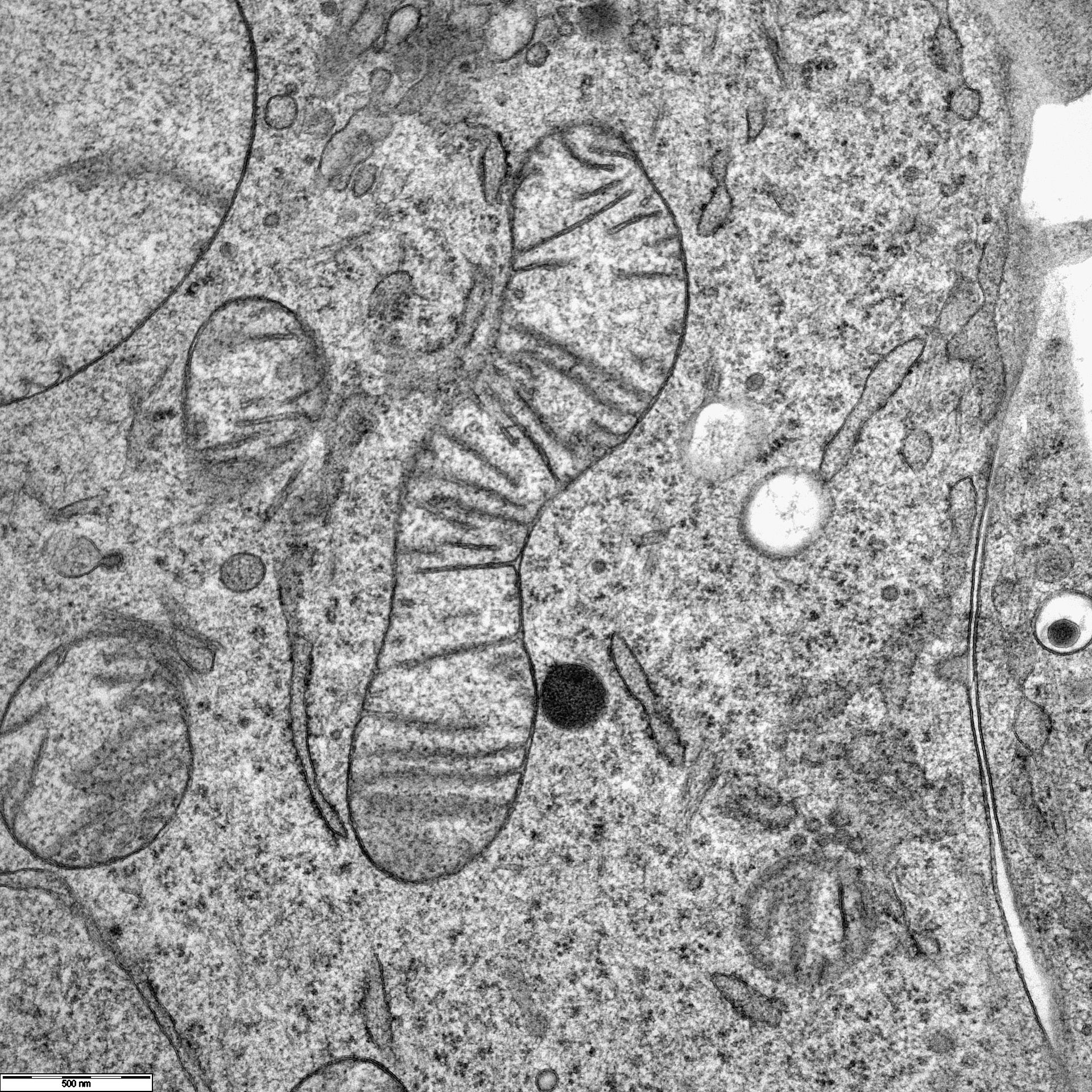
Unser Jeol 1400 TEM ist in Betrieb und wir sind begeistert von der Qualität der Bilder. Als Beispiel ein Mitochondrion aus einer Panc1 Zelle.
