Schematic representation of the antigen D on the erythrocyte´s
surface. The normal RhD protein is shown (left). Every sphere represents
an amino acid. Six extracellular loops of amino acids are shown (light yellow
and light grey). Transmembranous protein segments are depicted in reddish.
The RhD protein differs from the RhCE protein (not shown) be seven extracellular
amino acids only (yellow). All other extracellular amino acids (light yellow
and light blue) 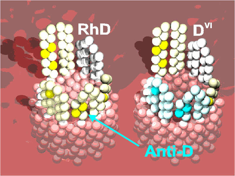 are identical between the RhD- and RhCE-protein. In the DVI-protein (right),
two loops carry the RhCE-specific sequence (light blue) rather than the RhD-specific
sequence. Thus, the extracellular parts of the DVI-protein differ from the
normal RhD-protein by three amino acids only (blue). Other transmembranous
and intracellular differences may, however, also effect the protein configuration
and are not shown for simplicity.
are identical between the RhD- and RhCE-protein. In the DVI-protein (right),
two loops carry the RhCE-specific sequence (light blue) rather than the RhD-specific
sequence. Thus, the extracellular parts of the DVI-protein differ from the
normal RhD-protein by three amino acids only (blue). Other transmembranous
and intracellular differences may, however, also effect the protein configuration
and are not shown for simplicity.